Lecture 5 – Looking through your skin
From the 1975 programme notes:
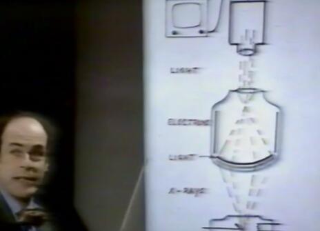
To be asked to diagnose a fault inside such a complex system as the human body without being able to look inside it would appear to make the task very difficult, yet this is a problem doctors had to face until the advent of X-rays at the turn of the century. Tissue and bone, except in very thin layers, are opaque to visible light; we cannot therefore look through or into the body in the ordinary sense at all. Other forms of electromagnetic radiation can penetrate tissue and bone to varying degrees, and with their aid a visible image can be reconstructed.
The most common form are X-rays which, until recently, were only capable of producing a shadow picture in which fine detail could be obscured by denser shadows. Recent developments in X-ray scanning and subsequent construction of the image point by point, with the aid of a computer, have produced a spectacular improvement.
Gamma-rays, of even shorter wavelength than X-rays, are emitted by certain radioactive isotopes. Some of these isotopes are taken up preferentially by particular tissues or organs when introduced into the body. The gamma-rays can pass through tissue and with appropriate equipment a crude image can be obtained which shows where in the body the isotope has accumulated.
Then there is infra-red radiation, of rather longer wavelength than light, which can penetrate tissue to a small extent. However it is also emitted from the skin surface in relation to its temperature, and therefore an infra-red picture of a portion of the body will really be a temperature map of the surface and structures just beneath it. This is useful because inflammation or abnormal rate of growth will result in high-temperature areas, and lack of blood supply in low-temperature regions.
Lastly there is ultrasound, not an electromagnetic radiation, but just a mechanical vibration in a material at a much higher frequency than ordinary sound. A beam of ultrasound aimed into the body will be partially reflected whenever it encounters a boundary between materials in which the velocity of sound is different. If the beam is pulsed one can measure how far away the reflecting boundary is by timing how long it takes for the echo to come back. If the beam is also moved to and fro across the surface, an image (in ultrasound terms) of a 'slice' of the patient can be obtained.
These methods enable us to see in many different ways, because the properties of the material which contribute to the image are different in every case.
About the 1975 CHRISTMAS LECTURES
In his 1975 CHRISTMAS LECTURES, Heinz Wolff explores how to investigate your inside without breaking the skin.
From the 1975 programme notes:
Imagine that you had to find out what was wrong with a motor car, but that you were not allowed to open the bonnet, or undo any nuts and bolts, or to break any wires. You would have to rely entirely on the noises you could hear, on how it reacted to the manipulation of external controls, and maybe on the characteristics of the exhaust.
The doctor using only his own senses is in much the same position when examining a patient, except that he has to deal with a very much more complicated system, and moreover one which is understood less completely than a motor car. This series of lectures is about how one can examine the functioning or the structure of the inside of the body noninvasively, that is, without having to open the patient. In particular, they will be concerned with the techniques which are now available to amplify the doctor's senses or to detect signals to which we are normally quite insensitive.
Each lecture will take a particular set of signals, consider their origin and why they are important, demonstrate how they are detected and measured, and explain how the instrumentation works. The lectures will also illustrate how much modern medicine is becoming dependent on a proper application and understanding of engineering and physical principles.
